TUMORS OF THE BRAIN AND SPINE
Remote patient diagnosis & consultation
To simplify the initial contact with our hospital for patients from abroad, we have prepared a remote two-stage process of prequalification for neurosurgical procedure. In the first stage, the patient sends via email all available medical documentation (lab results, treatment history, MRI scans) to be analyzed by our neurosurgical team. In second stage, we will arrange an on-line video consultation with one of our neurosurgeons. The cost of the online consultation is 200EUR. All necessary exams (e.g. CT, MR) and consultations will be repeated during the patient’s stay at Vital Medic Hospital. The final therapeutic decision will be based on neurosurgical exam and radiological results. At this point the final financial settlement may be presented to the patient. The neurosurgical treatment will be carried out according to the schedule determined by the team of specialists. We encourage you to use the email below or the contact form to begin your treatment.
Comprehensive and coordinated treatment
Comprehensive and coordinated treatment for brain and spine tumors takes place at the Brain and Spine Tumor Treatment Center, where we provide:
• Neurosurgical/neurological consultations – initial, to diagnose the disease
• Diagnostic tests appropriate for the initial diagnosis
• Qualification for surgical treatment
• Individually selected surgery with the use of advanced technology for planning and performing the procedure
• Individually planned perioperative and postoperative neurological rehabilitation
• Follow-up consultations for post-op control and continuation of the treatment
PROCEDURES
Glioma resection in Vital Medic
We perform surgery for high grade gliomas (known as glioblastomas), also for recurrent tumors.
We perform resections of brain gliomas located in:
• supra-tentorial region (all eloquent localizations)
• ventricular system
• insular lobe
• brainstem
• cerebellum
Neurosurgical removal of the tumor is performed as a first line treatment of the most of intercranial tumors. The goal of the surgery is to remove the tumor completely or, if this is not possible, to reduce the pathological mass as much as possible while maintaining neurological functions.
In our department we use up to date operational techniques to provide the maximal safety of resection and the optimal treatment results:
• Electrophysiological monitoring is used to intraoperative mapping of brain structures responsible for various neurological functions: movement, vision, etc.
• Neuropsychological monitoring is used during awake craniotomy to intraoperative mapping of brain structures responsible for cognitive functions, speech, memory, etc.
• Fluorescence microscopy to identify tumor margins
Treatment plan is based on a fusion of MR imaging with tractography, functional MRI of the brain tumor area and intraoperative tomography studies, using O-arm and neuronavigation.
Intraoperative Neuromonitoring
Intraoperative neuromonitoring is a diagnostic tool that is used during surgery. This method is based on the fact that nerve impulses are in the form of microelectric impulses which can be detected with super-sensitive equipment. During surgery, impulses are detected and recorded from the cortex of the brain responsible for example for hand movement. This method is also used for intraoperative mapping of eloquent structures for example visual or speech cortex. In that case direct electrical stimulation is used to induce neurological effects (speech arrest, visual disturbances, etc.) during awake craniotomy. This method allows to increase safety and improves neurological outcome of neurosurgical procedures. Intraoperative neuromonitoring performed during deep brain stimulation surgery is necessary for correct electrode placement by the neurosurgeon.
Intraoperative Fluorescence
Intraoperative fluorescence is an diagnostic tool based on changes in the metabolism of tumor cells that result in the presence of enzyme groups other than the typical ones. In this method drug is administered to the patient and it accumulates inside tumor cells. During surgery, the tumor region is illuminated with light of a specific wavelength, the areas infiltrated with tumor glow, making it easier to identify them. This helps the surgeon to perform complete resection and increase safety of the procedure. Fluorescence guided surgery is necessary in brain gliomas procedures to improve oncological outcome.
We perform resections of primary intracranial tumors (intrinsic) and secondary metastatic tumors (extrinsic).
The most commonly diagnosed intracranial tumors are gliomas originating from glial cells, meningiomas originating from arachnoid epithelial cells, and the rarest, neuroblastomas originating from cells of the peripheral nervous system.
Tumor resection is performed in locations:
• Subthalamic area (cerebellum, brainstem, great aperture area)
• Pineal region
• Cerebellopontine angle (CPA)
• Ventricular system
• Skull base and orbit
Neurosurgical removal of the tumor is used to treat tumors. The goal of the surgery is to remove the tumor completely or, if this is not possible, to reduce the tumor mass as much as possible while maintaining normal neurological function.
In Vital Medic Hospital during tumor resection surgery we use the most modern methods that bring optimal treatment results:
• Electrophysiological monitoring allows during surgery to identify brain structures responsible for vital functions: movement, speech, vision, etc.
• Neuropsychological monitoring during surgery helps identify brain structures responsible for cognitive functions and mood
• Treatment planning based on a fusion of MR imaging with tractography of the brain tumor area and intraoperative tomography studies, using O-arm and neuronavigation
• Fluorescence microscopy to identify tumor cells
Intraoperative Neuromonitoring
Intraoperative neuromonitoring is a diagnostic tool that is used during surgery. This method is based on the fact that nerve impulses are in the form of microelectric impulses which can be detected with super-sensitive equipment. During surgery, impulses are detected and recorded from the cortex of the brain responsible for example for hand movement. This method is also used for intraoperative mapping of eloquent structures for example visual or speech cortex. In that case direct electrical stimulation is used to induce neurological effects (speech arrest, visual disturbances, etc.) during awake craniotomy. This method allows to increase safety and improves neurological outcome of neurosurgical procedures. Intraoperative neuromonitoring performed during deep brain stimulation surgery is necessary for correct electrode placement by the neurosurgeon.
Intraoperative Fluorescence
Intraoperative fluorescence is an diagnostic tool based on changes in the metabolism of tumor cells that result in the presence of enzyme groups other than the typical ones. In this method drug is administered to the patient and it accumulates inside tumor cells. During surgery, the tumor region is illuminated with light of a specific wavelength, the areas infiltrated with tumor glow, making it easier to identify them. This helps the surgeon to perform complete resection and increase safety of the procedure. Fluorescence guided surgery is necessary in brain gliomas procedures to improve oncological outcome.
Biopsy is an invasive diagnostic method which allows to acquire sample of a lesion in non-operative cases. Tumor sample is histopathologically and genetically examined to obtain diagnosis and plan further treatment.
We perform:
• Stereotactic frame based biopsies
• Stereotactic frameless biopsies using neuronavigation
• Open biopsy procedures
• Fluorescent guided spinal biopsies
• Biopsies of intramedullary tumors
In our department we also can perform brain biopsies in children.
Cranioplasty is performed as a stand alone procedure or as an adjunct to neuro-oncologic surgery. In our department we use a standard cranioplasty implants or bespoke 3D implant dedicated to individual patient. The implants are made of polypropylene and polyester yarn and are characterized by high biocompatibility and resistance. We also perform cranioplasty using dedicated 3D implants made from hydroxyapatite or bone cement.
We perform resections of spinal column tumors and tumors located in spinal canal: epidural tumors, intradural – extramedullary tumors, intramedullary tumors. We also treat vascular diseases of the spinal cord (e.g. arteriovenous hemangiomas). Intraoperative neuromonitoring is used routinely during these procedures.
In non-operative cases, biopsy can be performed.
NEUROSURGERY DEPARTMENT
CONTACT
Vital Medic
46-200 Kluczbork
Skłodowskiej-Curie st. 21
Poland, opolskie region
Contact to our patient service:
📱 +48 531 999 172
📱 +48 531 439 671
✉ neuro@vitalmedic.pl
MAIN EMAIL
PATIENT COORDINATOR
Paweł Respondek
📞 +48 531 999 172
✉ p.respondek@vitalmedic.pl
PATIENT CARER
Nicoletta Nicowska
📞 +48 531 439 671
✉ n.nicowska@vitalmedic.pl
LEAVE US MESSAGE
WE WILL GET BACK TO YOU
PERSONNEL
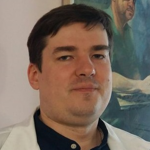
Witold Libionka, MD PhD
specialist in neurosurgery and neurotraumatology
Connected with neurosurgery since 1998, when he started his scientific activity and then, remaining a scholarship holder of the Ministry of Health, individual studies at the Department of Neurosurgery and Neurotraumatology of the Jagiellonian University Medical College. He continued his education at PhD studies at the Faculty of Medicine CMUJ in Krakow (2002-2006). During his specialisation in neurosurgery at the Department of Neurosurgery and Neurotraumatology of the Medical University of Krakow (2003-2010) he participated in numerous training courses to improve his professional qualifications (including Poland, USA, Germany, France). He completed a one-year research and clinical fellowship in functional and stereotactic neurosurgery at the Department of Neurosurgery, University of Rochester, USA.
Combining his professional and scientific work, he performs dozens of surgeries per month and participates in numerous research projects. He is the author or co-author of nearly a hundred publications and reports. His work on the mechanisms of action of deep brain stimulation has been awarded at national and international conferences and has been published in Nature Medicine and Nature Neuroscience.
He is an international consultant, teacher and lecturer in functional neurosurgery and oncology using functional mapping. He has expert experience in deep brain stimulation, spinal cord and peripheral nerve stimulation, intrathecal pump-assisted baclofen treatment, and resection of glial brain tumors in the eloquent area performed with intraoperative awakening and electrophysiological monitoring of speech, movement, vision, hearing, and tactile functions (more than a thousand such procedures performed in total).
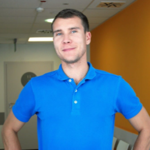
Bartosz Pańczyszak, Msc
physiotherapist
Manager of the rehabilitation facility at Vital Medic Hospital.
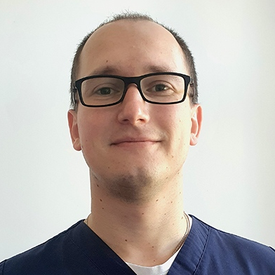
Mateusz Pawłowski, MD
specialist in neurosurgery
A graduate of the Faculty of Medicine in Zabrze, Silesian Medical University in Katowice. Participant of doctoral studies at the Medical University of Wroclaw. Doctoral student at the Medical University of Wroclaw.
Specializes in neurooncology, spine and hydrocephalus surgery. He has improved his knowledge and skills during numerous national and international courses.
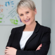
Anna Włodarczyk, Msc
psychologist, psychotherapist
- psychologist with clinical specialization, sexologist, psychotherapist
- certified instructor of addiction therapy. Graduate of the Warsaw School of Social Sciences and Humanities
- member of the Polish Psychological Association.
- court expert
- a participant of a specialized training for profilers of missing persons
- an expert of the Opole Province Headquarters of the State Fire Service in Opole in the area of widely understood psychological support
- implementer of preventive social campaigns, for which she was awarded the title “Man of the Year 2016 in the plebiscite of NTO”
- member of TSO, since 2006 closely associated with the State Fire Service as a Regional Rescue Specialist, in the field of psychological support for accident victims and firefighters
- trainer in the field of teamwork, team building, contact with victims, including the profiles of missing persons, debriefing and defusing
- handler of two dogs prepared for rescue work (with one she passed the exam of field specialty of class 0 of PSP)
- Psychologist in Vital Medic Hospital in Kluczbork
Professional interests: since the cooperation with the Vital Medic Hospital, neuropsychology comes to the fore (more on this soon) and associated with the love of dogs activity in the Foundation “Opolsar,” the unit specialized in the search for missing persons using rescue dogs.
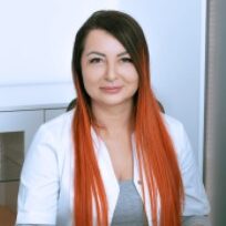
Monika Stomal-Słowińska, MD PhD
specialist in neurochirurgy
Monika Stomal-Slowinska, MD, graduated from the Silesian Medical Academy in Katowice in 1998. During her specialisation in neurosurgery she gained knowledge and improved her skills by participating in several trainings and courses abroad. Among others, she studied in Germany and Great Britain (London). In 2012, at the Medical University of Katowice, she was awarded the degree of Doctor of Medical Sciences for her thesis in neurosurgery. In 2015 she defended her PhD thesis in neuropsychology at the Faculty of Psychology, University of Warsaw. In the same year, she graduated with a degree in Aesthetic Medicine from the Medical College of Katowice, Poland, and a year later with a related degree in Anti-Aging Medicine from the International Center for Anti-Aging Medicine in Warsaw, a school under the auspices of the Society of Doctors of Aesthetic Dermatology (SLDE).
Monika Stomal-Slowinska, MD has many years of professional experience in the field of neurosurgery – she has been working in this field since her graduation.

Elżbieta Nowicka, MD PhD
specialist in clinical oncology and radiotherapy

Tamara Sokół, MD PhD
specialist in anaesthesiology and intensive care

Wojciech Fortuna, MD PhD
specialist in neurology
Professional experience:
- 2017-today – Department of Neurosurgery, Wroclaw Medical University – research and teaching fellow
- 2016-Today – Wrocław Walk Again Project – cell culture coordinator
- 2011-Today – Akson – Department of Therapeutic Rehabilitation of Spinal Cord Injuries and Diseases – neurological supervision over the course of neurorehabilitation, leading a program of neuromodulation of the nervous system using repetitive transcranial magnetic modulation (rTMS)
- 2011-2017 – Chair and Clinic of Neurosurgery, Medical University of Wrocław – scientific and technical specialist
- 2008-2011 – Institute of Immunology and Experimental Therapy, Polish Academy of Sciences, Wrocław – assistant professor, basic research, isolation and culture of human glial cells from nasal olfactory membrane used for transplantation to three patients with complete spinal cord injury
- 2008-2011 – Chair and Clinic of Neurology, Medical University of Wroclaw – specialization in neurology
- 2006-present – Medical Center, Center of Phage Therapy at the Institute of Immunology and Experimental Therapy of the Polish Academy of Sciences – physician, qualification of patients and conducting experimental therapy with bacteriophages
- 1997- present – Institute of Immunology and Experimental Therapy, Polish Academy of Sciences in Wrocław – assistant
- 1997-1999 – Department of Pediatrics, Immunology and Rheumatology, Medical University of Wrocław – specialization in pediatrics
- 1995-1996 – Center for Lung Diseases and Tuberculosis in Wrocław – resident physician, postgraduate internship
Achievements:
- Conducting electrophysiological intraoperative monitoring: micro- and macro-recording of brain activity during implantation of electrodes for deep brain stimulation (DBS), SEP sensory evoked potentials, MEP motor sensory evoked potentials, d-wave evaluation of D wave during spinal cord surgery, monitoring of cranial and peripheral nerves, mapping of cerebral cortex – in terms of movement, sensation and speech centers
- Conducting neuromodulation of the nervous system using repetitive transcranial magnetic modulation (rTMS) in patients with spinal cord injuries
- In vitro and in vivo cell research: establishment of primary cultures, cell culture and identification, assessment of cell biological properties
- Qualification and conduct of experimental bacteriophage therapy of chronic bacterial infections in humans

